원장칼럼
| 제목 | Ductal Carcinoma In-Situ (DCIS) [ 관상피내암] | 2018-06-15 |
|---|---|---|
|
https://blog.naver.com/gogngs/221299358368
관상피내암 은 생명을 위협하는 질환은 아니지만 경각심을 가져야 하는 병변 입니다. 관상피내암의 10년 생존률을 97%로 보고 하였습니다. 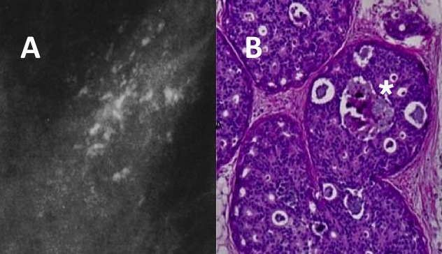 A: DCIS의 미세석회화 유방촬영소견 , B : 현미경사진 유방촬영은 DCIS의 정도를 과소 평가 할수 있습니다. 그래서 DCIS가 의심되는 모든 환자는 확대촬영을 통해 미세석회의 모양과 범위를 평가 해야 합니다. Classification of DCIS ( 관상피내암의 분류)관상피내암의 분류는 관내에서 비정상세포의 세포학적 형태와 자라는 양상에 따라 구분됩니다. 총 5개의 주요 형태로 분류합니다. The cribriform type of DCISback-to-back glands without intervening breast stroma. The epithelial cells in this subtype are typically small- to medium-sized, with uniform, dark cell nuclei. There are few mitoses and necrosis is only present in single cells or small cell clusters. The micro-papillary type of DCISsmall tufts of cells, oriented perpendicular to the basement membrane and projecting into the duct lumena. The apex of these projections can be broader than the base, giving a ‘club-shaped’ appearance. These intraluminal ‘micro-papillae’ do not have fibrovascular cores. The cells are usually small to medium in size, with dark cell nuclei and few mitoses. The papillary type of DCISintraluminal projections of tumor cells that do have fibrovascular cores. A variant of papillary DCIS is ‘intracystic papillary carcinoma’ which has tumor cells present in a single dilated space. The solid type of DCIStumor cells completely fill and distend the involved duct spaces, without necrosis, fenestrations or papillary projections. The tumor cells may be large, medium, or small. The ‘comedo’ type of DCIS has necrosis in the center of the involved duct spaces. This material is often calcified. These micro-calcifications may show up mammographically as linear orbranching calcifications. Tumor cells are large with nuclear pleomorphism and mitotic activity. The degree of ‘comedo necrosis’ in DCIS is a strong predictor for the risk of recurrence after treatment (Fisher et al., 1999). Grading of DCIS등급은 세포의 핵의 형태에 따라 나뉘어집니다. 그 기준들은 DCIS 의 크기와 형태, 세포 양극화, 미세석회이 존재와 위치, 괴사의 ㅣ존재와 형태 입니다. 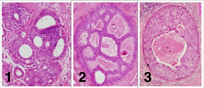 1. grade 1 2. grade 2 3. grade 3 DCIS 의 등급에따른 특별한 진단특징 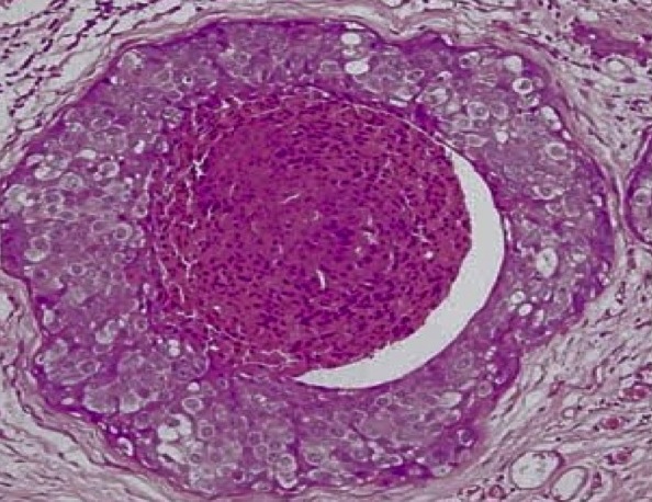 high grade comedo type DCIS Cancer Risk for DCIS ( 관상피내암의 암 위험도) | ||